-1.png)
.png)

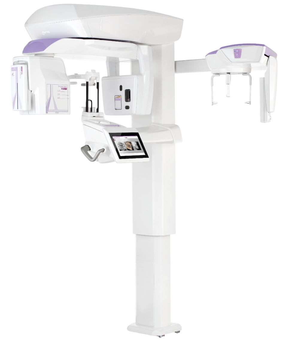
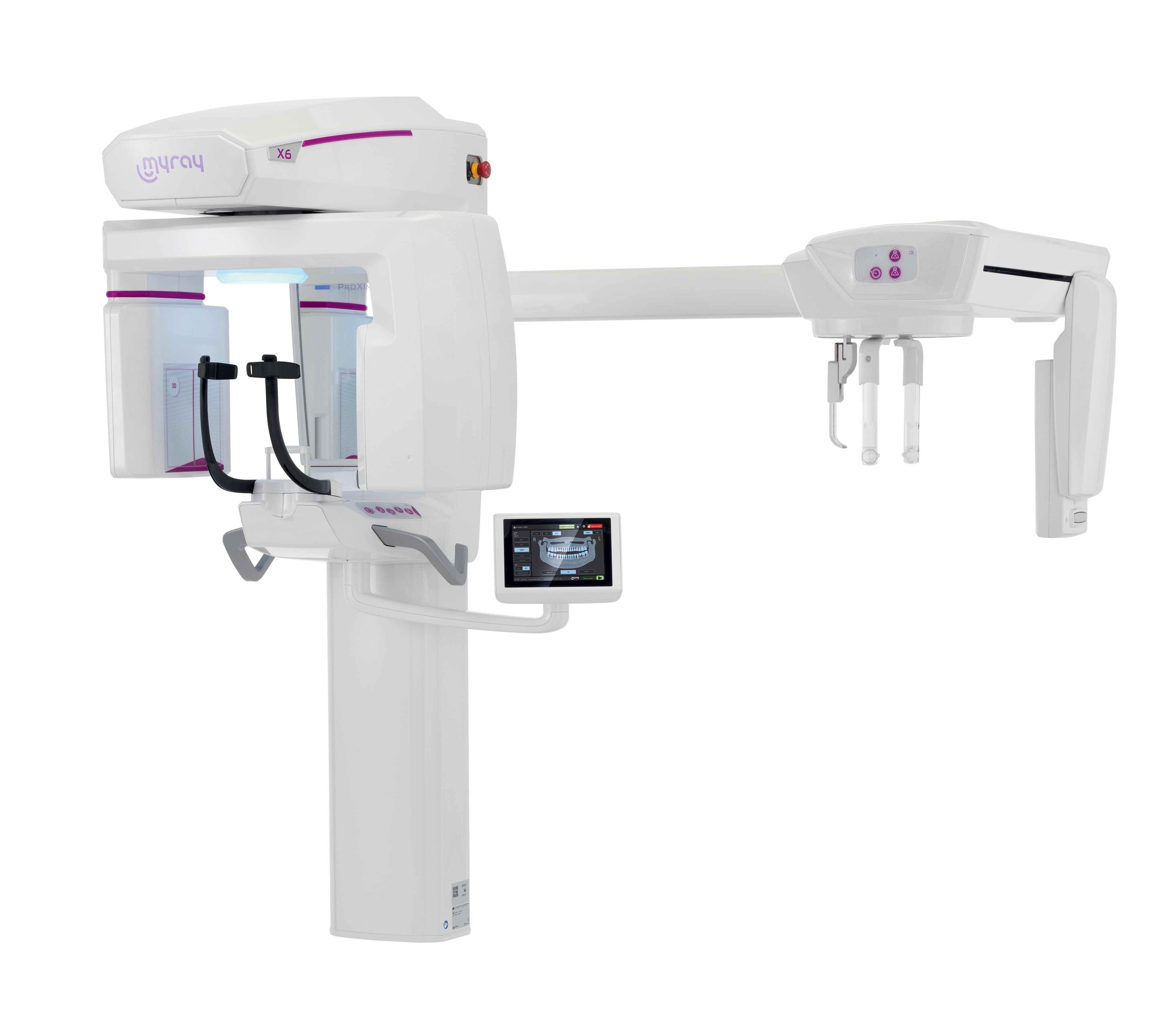
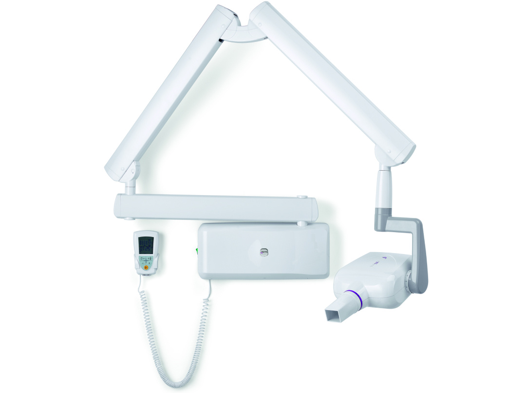
Extraoral
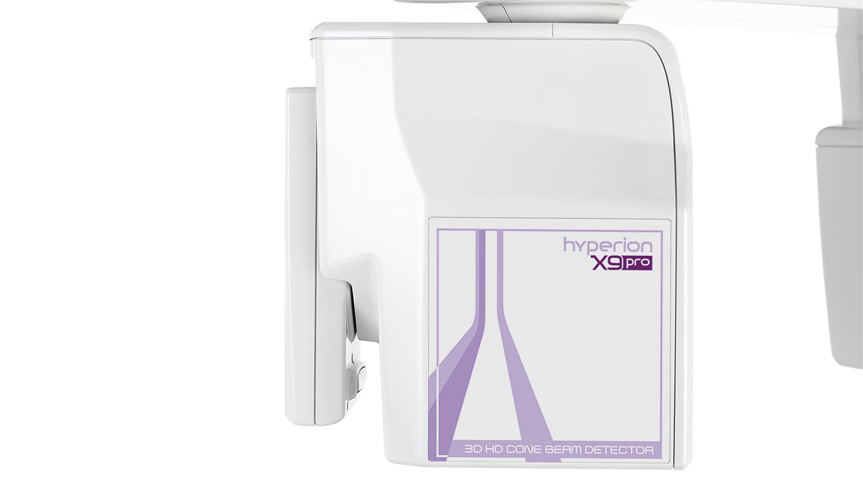
Hyperion X9 Pro
Designed for the future, built for you.
Characteristics
.png?width=1533&height=1080&name=Approvazione%20post%20(21).png)
Hyperion X9 Pro provides everything you need for advanced 2D, 3D and cephalometric diagnostic images. It delivers fast, safe, high-quality diagnoses, tailored to your needs at all times.
Workflow
With Hyperion X9 Pro practitioners can count on an optimised workflow that saves time and improves quality of service.
With iPad control, you can conveniently manage scans and settings via a tablet, with the user-friendly interface providing quick access to all the key functions.
The virtual control panel for PC allows comprehensive management of images/configurations and precise control of each stage of the process.
Full Touch control panel
A 10’’ full-touch control panel, positioned directly on the machine, allows fast selection of pre-set programmes and optimises the workflow.
The Face to Face system and the laser beams guide you towards correct patient positioning with ease, ensuring precise, comfortable scans.

Interactive View FOV
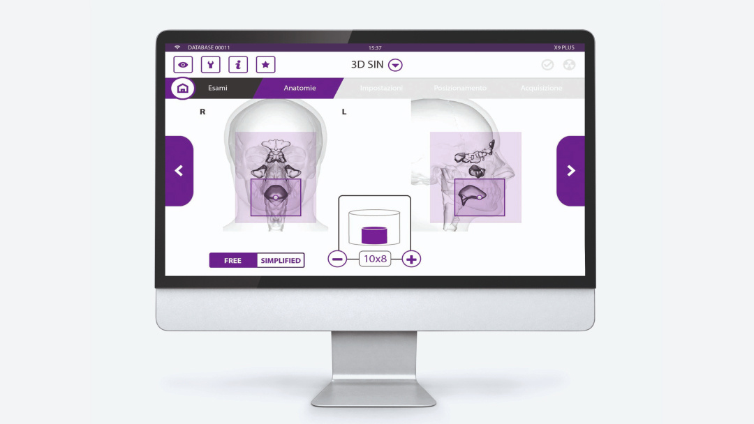
The Interactive View FOV option lets you redefine the size and position of the scan area directly on the patient's face, acquired frontally and laterally with 2 cameras using the on-machine control panel.
The Scout View system lets you align the volume on the area of interest directly from the PC. You can thus display two images (sagittal and frontal), obtained with ultra-low emissions, and modify the scan area with precision.
Thanks to the integrated software you can manage, process and share images quickly and easily, merging them into the clinical workflow with ease.
Capture every detail
Hyperion X9 Pro provides advanced 2D, 3D and cephalometric diagnostic images. Its modular configuration seamlessly adapts to your practice.
Direct conversion sensor
The DC''' direct-conversion sensor provides ultra-low dose SuperHD images by converting X-rays directly into electrical signals. Powerful Image Enhancer (PiE) filters optimise every detail, even on fast scans such as QuickPAN and QuickCEPH.

MultiPAN function
The MultiPAN function lets you generate from 5 (with a standard sensor) to 11 focus layers in a single scan with a direct conversion sensor; afterwards, simply select the one that best suits each diagnostic need.
The PAN scan uses MRT (Morphology Recognition Technology) and the iPAN protocol to automatically generate high-quality panoramic images. A state-of-the-art algorithm selects the best focus.

Laser tracing
The Face to Face system and laser tracing ensure fast, precise, comfortable positioning.

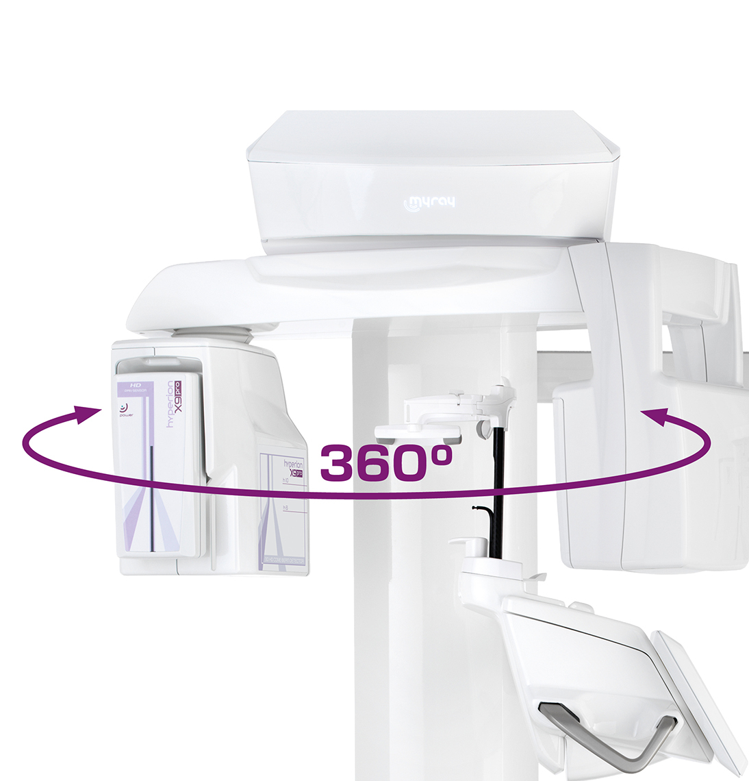
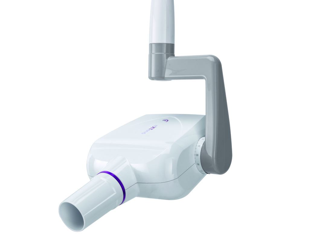
Ceph arm
The compact, reversible cephalometric arm easily adapts to your needs to provide ultra-fast or high-quality scanning options.

.png "Banner download MyRay (5)")
Filter by typology
-1.png)
-1.png)
-1.png)
-1.png)
-1.png)
.png)
-1.png)
-1.png)
-1.png)
